202 Tribble Gap Road, Suite 101, Cumming, GA 30040
We speak English, Spanish and Portuguese
We speak English, Spanish and Portuguese

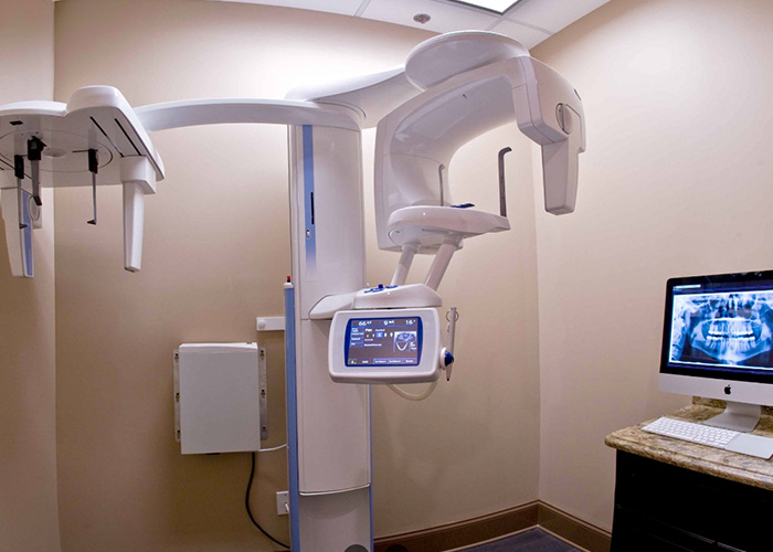
Digital radiography replaces traditional film with sensitive electronic sensors and software to capture and display dental images. Instead of exposing film to X‑ray photons and processing it with chemicals, the sensor converts X‑ray energy into a digital signal that the computer interprets as a high‑resolution image. The result is an image that appears on a monitor almost instantly, allowing clinicians to evaluate dental anatomy, restorations, and bone structures without delay. This technological shift has transformed how dental teams collect diagnostic information and communicate treatment needs.
The workflow for digital radiography is straightforward: a small sensor is positioned in the mouth or an external panel is used for extraoral views, then a digital capture device records the exposure. The captured image is sent immediately to the practice’s imaging software where it is stored in the patient record and can be enhanced, measured, or annotated. Because the image is digital from the outset, clinicians can apply tools such as contrast adjustment, magnification, and measurement overlays to reveal details that may be hard to see on film.
For patients, understanding this process helps demystify what happens during routine X‑rays. The equipment is compact and designed for minimal discomfort, and views that once required waiting for film development are now available within seconds. At Inspirational Smiles we use digital radiography to support faster, more informed conversations between patients and clinicians while maintaining the precision needed for accurate diagnosis and treatment planning.
One of the primary advantages of digital radiography is a marked reduction in radiation exposure compared with conventional film techniques. Digital sensors require less X‑ray energy to produce a usable image, and modern X‑ray units are engineered with focused beams and shielding to further limit exposure. These improvements align with the principle of ALARA — keeping radiation "As Low As Reasonably Achievable" — which guides responsible imaging practices in dentistry and medicine.
Beyond lower doses, digital systems enable clinicians to capture exactly the views they need without repeat exposures caused by poor film placement or processing errors. Because images are available immediately, the dental team can confirm adequacy of the image in real time and make adjustments on the spot rather than asking the patient to return for retakes. This immediacy reduces cumulative exposure over time, particularly important for patients who require periodic monitoring or complex treatment sequences.
Safety also extends to infection control and equipment handling. Digital sensors are designed with durable, easy‑to‑clean surfaces and can be covered with disposable barriers when placed intraorally. These practical protections help ensure imaging is both safe and hygienic for patients of all ages while supporting a streamlined clinical workflow.
The diagnostic value of digital radiography goes beyond exposure reduction; image quality and post‑capture manipulation are powerful clinical assets. High‑resolution sensors capture fine anatomic detail, and digital software allows for enhancement techniques—such as contrast, sharpening, and edge detection—that can make subtle features more visible. These tools help clinicians detect early decay between teeth, identify hairline root fractures, evaluate bone levels around teeth and implants, and assess the fit of restorations with greater confidence.
Because digital images can be enlarged without degradation, clinicians can examine areas of interest at a level of detail that film cannot match. Measurement tools built into imaging software provide precise numbers for root lengths, bone height, and implant planning dimensions, supporting decisions that rely on accurate, quantifiable data. This objective information helps align clinical assessments across the care team and supports consistent treatment outcomes.
Digital radiography also facilitates longitudinal comparison. Images taken at different visits can be displayed side‑by‑side, allowing the clinician to track changes in tooth structure, bone density, or restoration integrity over time. These comparative views are invaluable for monitoring healing, evaluating disease progression, and documenting the effectiveness of periodontal or endodontic therapies.
One of the lesser‑seen but highly practical benefits of digital imaging is how well it integrates into a modern dental practice’s software ecosystem. Digital radiographs are saved directly into the electronic health record (EHR), where they become part of the patient chart and can be associated with notes, treatment plans, and referral documentation. This centralized approach reduces paperwork, minimizes the risk of misplaced films, and streamlines communication between team members.
When collaboration is needed — for example, with a specialist, laboratory, or another dental office — digital files can be shared quickly and securely. High‑quality images can be exported in standard formats and sent over encrypted channels to preserve patient privacy while enabling consultative review. The ability to transmit images electronically shortens turnaround times for specialist input and supports coordinated care for complex cases such as implant planning or orthodontic treatment.
Data governance is an important consideration. Digital images are subject to the same privacy and security expectations as other health records, so practices implement access controls, regular backups, and secure storage protocols. These measures protect patient information while ensuring that diagnostic images remain available when needed for ongoing care.
From a patient standpoint, digital radiography enhances comfort and convenience. Sensors are slim and ergonomically designed to reduce gagging and discomfort during intraoral exposures, and the quick capture time means less time spent holding a sensor in place. Immediate image viewing also allows the clinician to involve the patient in the diagnostic process by showing findings on the monitor and explaining treatment rationale using visual evidence.
Digital imaging workflows often shorten appointment length because there is no waiting for film processing. This efficiency can make visits smoother and more predictable for patients with busy schedules or those who are anxious about long procedures. For children, seniors, and patients with special needs, the speed and clarity of digital images can make routine imaging less stressful and more tolerable.
There are environmental advantages as well: digital radiography eliminates the need for chemical developers, fixers, and paper handling associated with film processing. Removing these consumables reduces hazardous waste and aligns dental practices with broader sustainability goals. Taken together, these practical, comfort, and environmental factors contribute to a modern, patient‑centered approach to dental imaging.
In summary, digital radiography is a safer, faster, and more flexible alternative to traditional film X‑rays that supports accurate diagnosis, efficient clinical workflows, and improved patient experience. At Inspirational Smiles we use advanced digital imaging to inform treatment planning, enhance communication, and maintain high standards of safety and data security. If you have questions about how digital radiography is used in our office or how it might apply to your care, please contact us for more information.


Digital radiography is a modern alternative to traditional dental X-rays. It uses electronic sensors instead of film to capture high-resolution images of your teeth and jaw, which appear instantly on a computer screen. This allows your dentist to evaluate your oral health quickly and accurately.
Yes. Digital X-rays use significantly less radiation than traditional film X-rays — often up to 80% less — and follow the ALARA principle (As Low As Reasonably Achievable) to ensure maximum patient safety. Sensors are also designed with protective coverings and are fully sanitized between patients.
The process is simple and comfortable. A small digital sensor is placed in your mouth, or an external device is positioned for panoramic or extraoral images. The image is captured in seconds and sent immediately to the dentist’s computer for review, without any waiting time for film development.
Digital radiography provides faster results, sharper images, and the ability to enhance, zoom, and measure areas for better diagnosis. It also reduces the need for retakes, minimizes patient exposure, and eliminates harmful chemicals used in traditional film processing — making it safer and more eco-friendly.
Because digital images are stored electronically, they can be easily compared with previous X-rays to track changes in your oral health over time. They can also be securely shared with specialists, used for implant or orthodontic planning, and stored in your electronic health record for future reference.
Looking to schedule your next dental visit or learn more about our services?
Getting in touch with Inspirational Smiles is simple! Our caring team is ready to help with appointment scheduling, questions about treatments, or any concerns you may have. You can call us or use our easy online contact form—whatever works best for you.
Take the first step toward a healthier, more confident smile today and experience the difference that personalized, compassionate dental care can make.