202 Tribble Gap Road, Suite 101, Cumming, GA 30040
We speak English, Spanish and Portuguese
We speak English, Spanish and Portuguese

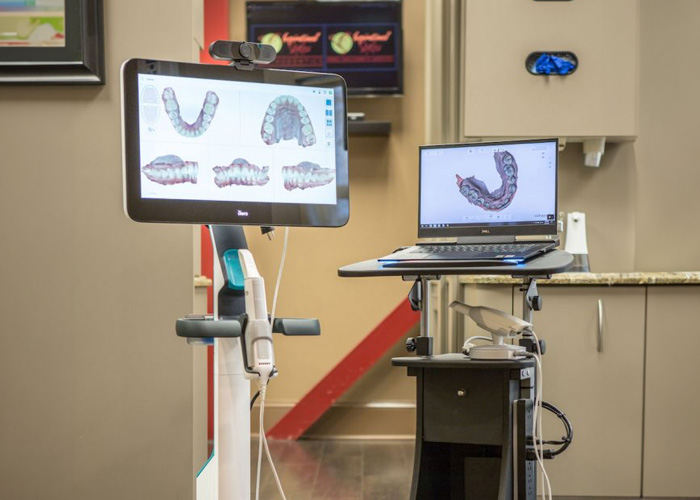
An intraoral camera is a compact, pen-sized imaging tool that captures high-resolution, full-color photos of the teeth, gums, and other soft tissues inside the mouth. Unlike a visual exam alone, these images reveal small fractures, early decay, worn restorations, and subtle changes in soft tissue that can be difficult to spot with the naked eye. For patients, the result is a clearer picture of oral health—literally and figuratively.
Because the camera records detailed images in real time, clinicians can examine suspicious areas from multiple angles and magnifications. This means minor problems are more likely to be detected early, when treatment can be simpler and less invasive. The level of detail also supports more precise diagnosis and targeted care planning.
For people concerned about oral cancer, gum disease, or unexplained symptoms, intraoral imaging can provide tangible evidence to guide next steps. It does not replace other diagnostic tools such as x-rays or clinical probing, but it complements them by offering a surface-level, color-accurate view that enhances overall assessment.
Patients often find that seeing a clear image of the issue helps them understand their condition more quickly. Visual information reduces uncertainty and makes the conversation about treatment options more productive and collaborative.
One of the biggest advantages of intraoral cameras is how they transform communication between clinicians and patients. Instead of relying solely on verbal descriptions, dentists can point to an on-screen image and explain what they see, why it matters, and which options are available. This visual approach helps patients make informed decisions about their care.
Images captured with the camera can be annotated and saved to the patient record, so they become part of a clear, shared narrative about oral health over time. During follow-up visits, clinicians can pull up prior images to demonstrate improvement, progression, or the effects of a chosen treatment—making the planning process transparent and evidence-based.
For interdisciplinary cases, intraoral photos are a practical way to share findings with specialists, hygienists, and dental laboratories. Clear, standardized images reduce the chance of miscommunication and ensure everyone involved has the same visual reference when discussing restorations, orthodontic work, or periodontal therapy.
Clinicians also use intraoral imaging to document treatment needs for administrative tasks such as insurance submissions or preauthorization requests. While not a substitute for formal diagnostic tools, these images add clarity to clinical reports and streamline many workflow steps behind the scenes.
Using an intraoral camera is quick and noninvasive. During a routine exam, the clinician or hygienist will gently guide the camera around the mouth while you remain seated in the dental chair. The device captures multiple views in a matter of seconds, and images appear instantly on a nearby monitor for joint review.
Patients typically experience no discomfort; the camera’s small size and ergonomic shape make it easy to position in tight areas without triggering a gag reflex. The process is safe for people of all ages, including children and seniors, and it generally adds only a few minutes to a standard check-up.
Because images are visible in real time, patients can ask questions and receive immediate explanations about what they are seeing. This interactive element often reduces anxiety and builds trust, especially when treatment recommendations are based on clearly visible evidence rather than abstract descriptions.
If additional imaging or diagnostic tests are needed, the intraoral camera images help determine which steps are most appropriate. In many cases, the camera clarifies whether a small concern can be monitored or if prompt intervention is prudent.
Intraoral photos are saved directly into a patient’s chart and become part of the permanent dental record. This systematic documentation creates a visual timeline that supports continuity of care and helps clinicians track changes across visits. A photographic record is particularly useful for monitoring restorations, checking for recurrent decay, and evaluating tissue healing after procedures.
When patients are referred to specialists or require laboratory-fabricated restorations, saved images can be shared to provide a clear, visual context for the work to be done. This reduces guesswork and improves the fit, function, and aesthetics of final restorations by giving lab technicians and specialists a precise reference.
Saved intraoral images are also valuable for risk management and informed consent. When treatment recommendations are accompanied by visual documentation, both patients and clinicians have a transparent basis for decisions and follow-up plans. This level of record-keeping supports safer, more accountable care.
Patients retain the option to request copies of their images for personal records or for transfer to another provider. Having this visual history available can simplify future transitions and make second opinions more efficient when they are sought.
Modern intraoral cameras use safe, low-energy LED lighting and high-resolution sensors designed specifically for dental use. They do not emit ionizing radiation and are therefore considered a safe complement to radiographic imaging. The devices are built for repeated clinical use and are subject to sterilization and infection-control protocols between patients.
Designers of intraoral cameras focus on comfort and maneuverability: curved tips, slim profiles, and anti-glare lighting minimize patient discomfort while providing consistently clear images. The software that accompanies these systems often includes tools for zooming, measuring, and comparing images side-by-side, which improves diagnostic accuracy and patient education.
Clinics integrate intraoral camera systems with practice management and imaging software so photos are stored, retrieved, and displayed efficiently. This interoperability reduces administrative friction and ensures that images are available when needed for treatment planning, follow-up, or interdisciplinary care.
As imaging technology continues to evolve, intraoral cameras remain an accessible, cost-effective way to bring high-quality visuals into routine dental care. Their role is not to replace other diagnostic modalities but to complement them—adding a layer of clarity that benefits both patients and clinicians.
At Inspirational Smiles in Cumming, GA, our team uses intraoral imaging to ensure patients see and understand the factors behind recommended care. If you have questions about intraoral cameras or want to learn how these images could be used in your treatment, please contact us for more information.


An intraoral camera is a small, pen-sized device that captures high-resolution, full-color photos of the inside of your mouth. The images are displayed instantly on a monitor, allowing you and your dentist to see teeth, gums, and soft tissues in real time.
Intraoral cameras help detect issues early by revealing small cracks, early decay, worn fillings, and soft tissue changes that are hard to see with the naked eye. They support more accurate diagnosis and targeted treatment planning.
No. The camera is very small and ergonomically designed for patient comfort. Most people experience no discomfort, even when images are taken of hard-to-reach areas. The process is quick and noninvasive, adding only a few minutes to your appointment.
Not exactly. Intraoral cameras provide detailed surface images, while X-rays show structures below the surface like bone and tooth roots. Used together, they give a more complete picture of your oral health.
Yes. Intraoral images are stored in your digital dental record and can be securely shared with specialists, labs, or other dental offices if needed. This ensures continuity of care and helps improve treatment outcomes.
Looking to schedule your next dental visit or learn more about our services?
Getting in touch with Inspirational Smiles is simple! Our caring team is ready to help with appointment scheduling, questions about treatments, or any concerns you may have. You can call us or use our easy online contact form—whatever works best for you.
Take the first step toward a healthier, more confident smile today and experience the difference that personalized, compassionate dental care can make.